A dentist can see three of the five surfaces of a back tooth. The two that touch the neighbouring teeth are hidden, and they are a common place for decay to start.
X-rays exist to look at the parts of your mouth that light cannot reach.
What they show
Bitewings — the ones you bite a tab for — show the crowns of the back teeth on both jaws, and are the standard way of finding decay between teeth and checking the bone level at the gum.
Periapicals show a whole tooth including the root tip, and are used when a specific tooth is symptomatic, before extraction, and during root canal treatment.
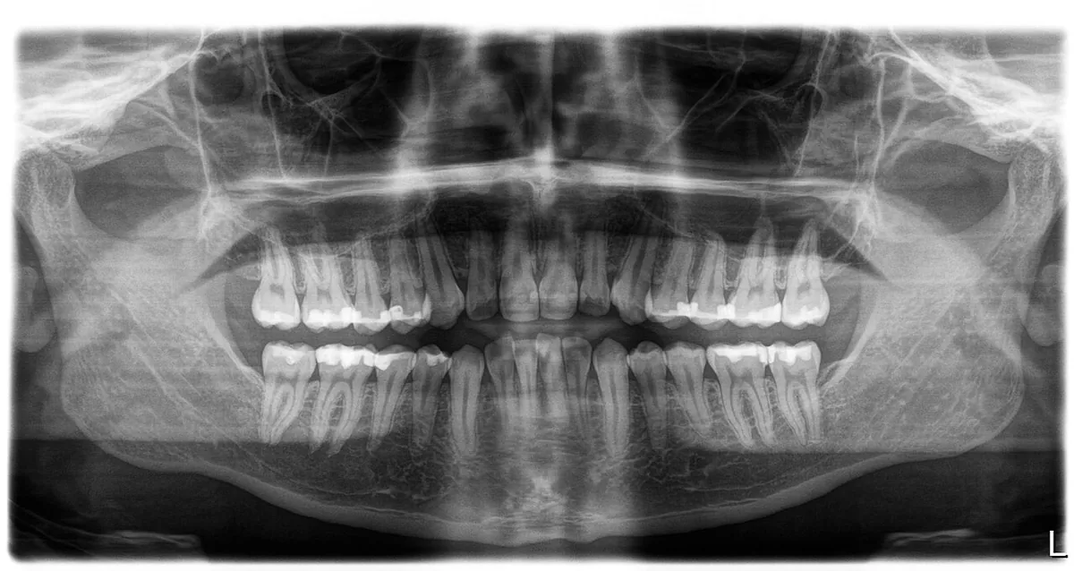
Panoramic (OPG) images sweep around the head to show both jaws, the sinuses and the jaw joints. These are used for wisdom teeth, trauma, implant planning and anything affecting the jaw as a whole.
The dose question
This is the part people actually want answered, so plainly: dental radiography uses very small doses. A set of bitewings is a tiny fraction of what you receive from natural background radiation in a year — the air, the ground and the food around you.
That does not make it nothing, which is why the professional standard is to justify every image individually. An X-ray should be taken because a specific question needs answering, not because it is that time of year.
You should be able to ask what a particular image is for and get a specific answer.
How often
Intervals are set by risk, not by the calendar:
- Someone with repeated new decay may need bitewings every six to twelve months
- Someone with a stable mouth and no history may go two years or considerably longer
- Children’s intervals differ again, because decay progresses faster through thinner enamel
Your risk changes. An interval that was right five years ago may not be right now, in either direction.
If you are pregnant
Dental X-rays are not directed at the abdomen and the doses involved are extremely small. UK guidance is that the risk to a developing baby is negligible, which is why you may not even be asked about pregnancy before a dental X-ray. Being pregnant is not a reason a dental X-ray cannot be taken. If an image is not needed right now it can of course wait — but that is true whether or not you are pregnant.
You will not be given a lead apron, and that is deliberate rather than an oversight. UK guidance is that there is no justification for using aprons routinely in dental radiography: the beam never points at the abdomen, so the dose reaching it is negligible, and what little scatter there is happens inside the body, where an apron on the outside cannot do anything. A thyroid collar is the same — not routine, but it is used for the few views where the beam does pass close to the thyroid. Tell your dentist you are pregnant regardless.
What it is like
Modern sensors are digital, so the image appears immediately and the exposure is a fraction of a second. The awkward part is purely mechanical: holding a sensor against the roof of your mouth can trigger a gag reflex.
If you gag easily, say so beforehand. Smaller sensors, a different holder, sitting more upright, and breathing through your nose all help, and a panoramic image avoids putting anything inside the mouth at all.
The point of an X-ray is not the picture. It is finding the thing while it is still small enough to be a small problem.